
4.1: Tipos de Tejidos
- Page ID
- 123210

\( \newcommand{\vecs}[1]{\overset { \scriptstyle \rightharpoonup} {\mathbf{#1}} } \)
\( \newcommand{\vecd}[1]{\overset{-\!-\!\rightharpoonup}{\vphantom{a}\smash {#1}}} \)
\( \newcommand{\id}{\mathrm{id}}\) \( \newcommand{\Span}{\mathrm{span}}\)
( \newcommand{\kernel}{\mathrm{null}\,}\) \( \newcommand{\range}{\mathrm{range}\,}\)
\( \newcommand{\RealPart}{\mathrm{Re}}\) \( \newcommand{\ImaginaryPart}{\mathrm{Im}}\)
\( \newcommand{\Argument}{\mathrm{Arg}}\) \( \newcommand{\norm}[1]{\| #1 \|}\)
\( \newcommand{\inner}[2]{\langle #1, #2 \rangle}\)
\( \newcommand{\Span}{\mathrm{span}}\)
\( \newcommand{\id}{\mathrm{id}}\)
\( \newcommand{\Span}{\mathrm{span}}\)
\( \newcommand{\kernel}{\mathrm{null}\,}\)
\( \newcommand{\range}{\mathrm{range}\,}\)
\( \newcommand{\RealPart}{\mathrm{Re}}\)
\( \newcommand{\ImaginaryPart}{\mathrm{Im}}\)
\( \newcommand{\Argument}{\mathrm{Arg}}\)
\( \newcommand{\norm}[1]{\| #1 \|}\)
\( \newcommand{\inner}[2]{\langle #1, #2 \rangle}\)
\( \newcommand{\Span}{\mathrm{span}}\) \( \newcommand{\AA}{\unicode[.8,0]{x212B}}\)
\( \newcommand{\vectorA}[1]{\vec{#1}} % arrow\)
\( \newcommand{\vectorAt}[1]{\vec{\text{#1}}} % arrow\)
\( \newcommand{\vectorB}[1]{\overset { \scriptstyle \rightharpoonup} {\mathbf{#1}} } \)
\( \newcommand{\vectorC}[1]{\textbf{#1}} \)
\( \newcommand{\vectorD}[1]{\overrightarrow{#1}} \)
\( \newcommand{\vectorDt}[1]{\overrightarrow{\text{#1}}} \)
\( \newcommand{\vectE}[1]{\overset{-\!-\!\rightharpoonup}{\vphantom{a}\smash{\mathbf {#1}}}} \)
\( \newcommand{\vecs}[1]{\overset { \scriptstyle \rightharpoonup} {\mathbf{#1}} } \)
\( \newcommand{\vecd}[1]{\overset{-\!-\!\rightharpoonup}{\vphantom{a}\smash {#1}}} \)
\(\newcommand{\avec}{\mathbf a}\) \(\newcommand{\bvec}{\mathbf b}\) \(\newcommand{\cvec}{\mathbf c}\) \(\newcommand{\dvec}{\mathbf d}\) \(\newcommand{\dtil}{\widetilde{\mathbf d}}\) \(\newcommand{\evec}{\mathbf e}\) \(\newcommand{\fvec}{\mathbf f}\) \(\newcommand{\nvec}{\mathbf n}\) \(\newcommand{\pvec}{\mathbf p}\) \(\newcommand{\qvec}{\mathbf q}\) \(\newcommand{\svec}{\mathbf s}\) \(\newcommand{\tvec}{\mathbf t}\) \(\newcommand{\uvec}{\mathbf u}\) \(\newcommand{\vvec}{\mathbf v}\) \(\newcommand{\wvec}{\mathbf w}\) \(\newcommand{\xvec}{\mathbf x}\) \(\newcommand{\yvec}{\mathbf y}\) \(\newcommand{\zvec}{\mathbf z}\) \(\newcommand{\rvec}{\mathbf r}\) \(\newcommand{\mvec}{\mathbf m}\) \(\newcommand{\zerovec}{\mathbf 0}\) \(\newcommand{\onevec}{\mathbf 1}\) \(\newcommand{\real}{\mathbb R}\) \(\newcommand{\twovec}[2]{\left[\begin{array}{r}#1 \\ #2 \end{array}\right]}\) \(\newcommand{\ctwovec}[2]{\left[\begin{array}{c}#1 \\ #2 \end{array}\right]}\) \(\newcommand{\threevec}[3]{\left[\begin{array}{r}#1 \\ #2 \\ #3 \end{array}\right]}\) \(\newcommand{\cthreevec}[3]{\left[\begin{array}{c}#1 \\ #2 \\ #3 \end{array}\right]}\) \(\newcommand{\fourvec}[4]{\left[\begin{array}{r}#1 \\ #2 \\ #3 \\ #4 \end{array}\right]}\) \(\newcommand{\cfourvec}[4]{\left[\begin{array}{c}#1 \\ #2 \\ #3 \\ #4 \end{array}\right]}\) \(\newcommand{\fivevec}[5]{\left[\begin{array}{r}#1 \\ #2 \\ #3 \\ #4 \\ #5 \\ \end{array}\right]}\) \(\newcommand{\cfivevec}[5]{\left[\begin{array}{c}#1 \\ #2 \\ #3 \\ #4 \\ #5 \\ \end{array}\right]}\) \(\newcommand{\mattwo}[4]{\left[\begin{array}{rr}#1 \amp #2 \\ #3 \amp #4 \\ \end{array}\right]}\) \(\newcommand{\laspan}[1]{\text{Span}\{#1\}}\) \(\newcommand{\bcal}{\cal B}\) \(\newcommand{\ccal}{\cal C}\) \(\newcommand{\scal}{\cal S}\) \(\newcommand{\wcal}{\cal W}\) \(\newcommand{\ecal}{\cal E}\) \(\newcommand{\coords}[2]{\left\{#1\right\}_{#2}}\) \(\newcommand{\gray}[1]{\color{gray}{#1}}\) \(\newcommand{\lgray}[1]{\color{lightgray}{#1}}\) \(\newcommand{\rank}{\operatorname{rank}}\) \(\newcommand{\row}{\text{Row}}\) \(\newcommand{\col}{\text{Col}}\) \(\renewcommand{\row}{\text{Row}}\) \(\newcommand{\nul}{\text{Nul}}\) \(\newcommand{\var}{\text{Var}}\) \(\newcommand{\corr}{\text{corr}}\) \(\newcommand{\len}[1]{\left|#1\right|}\) \(\newcommand{\bbar}{\overline{\bvec}}\) \(\newcommand{\bhat}{\widehat{\bvec}}\) \(\newcommand{\bperp}{\bvec^\perp}\) \(\newcommand{\xhat}{\widehat{\xvec}}\) \(\newcommand{\vhat}{\widehat{\vvec}}\) \(\newcommand{\uhat}{\widehat{\uvec}}\) \(\newcommand{\what}{\widehat{\wvec}}\) \(\newcommand{\Sighat}{\widehat{\Sigma}}\) \(\newcommand{\lt}{<}\) \(\newcommand{\gt}{>}\) \(\newcommand{\amp}{&}\) \(\definecolor{fillinmathshade}{gray}{0.9}\)8Objetivos de aprendizaje
- Identificar los cuatro tipos principales de tejido
- Discutir las funciones de cada tipo de tejido
- Relacionar la estructura de cada tipo de tejido con su función
- Discutir el origen embrionario del tejido
- Identificar las tres capas germinales principales
- Identificar los principales tipos de membranas tisulares
El término tejido se utiliza para describir un grupo de células que se encuentran juntas en el cuerpo. Las células dentro de un tejido comparten un origen embrionario común. La observación microscópica revela que las células en un tejido comparten características morfológicas y están dispuestas en un patrón ordenado que logra las funciones del tejido. Desde la perspectiva evolutiva, los tejidos aparecen en organismos más complejos. Por ejemplo, los protistas multicelulares, eucariotas antiguos, no tienen células organizadas en tejidos.
Aunque existen muchos tipos de células en el cuerpo humano, están organizadas en cuatro amplias categorías de tejidos: epiteliales, conectivos, musculares y nerviosos. Cada una de estas categorías se caracteriza por funciones específicas que contribuyen a la salud general y al mantenimiento del cuerpo. Una alteración de la estructura es un signo de lesión o enfermedad. Dichos cambios se pueden detectar a través de la histología, el estudio microscópico de la apariencia, organización y función de los tejidos.
Los cuatro tipos de tejidos
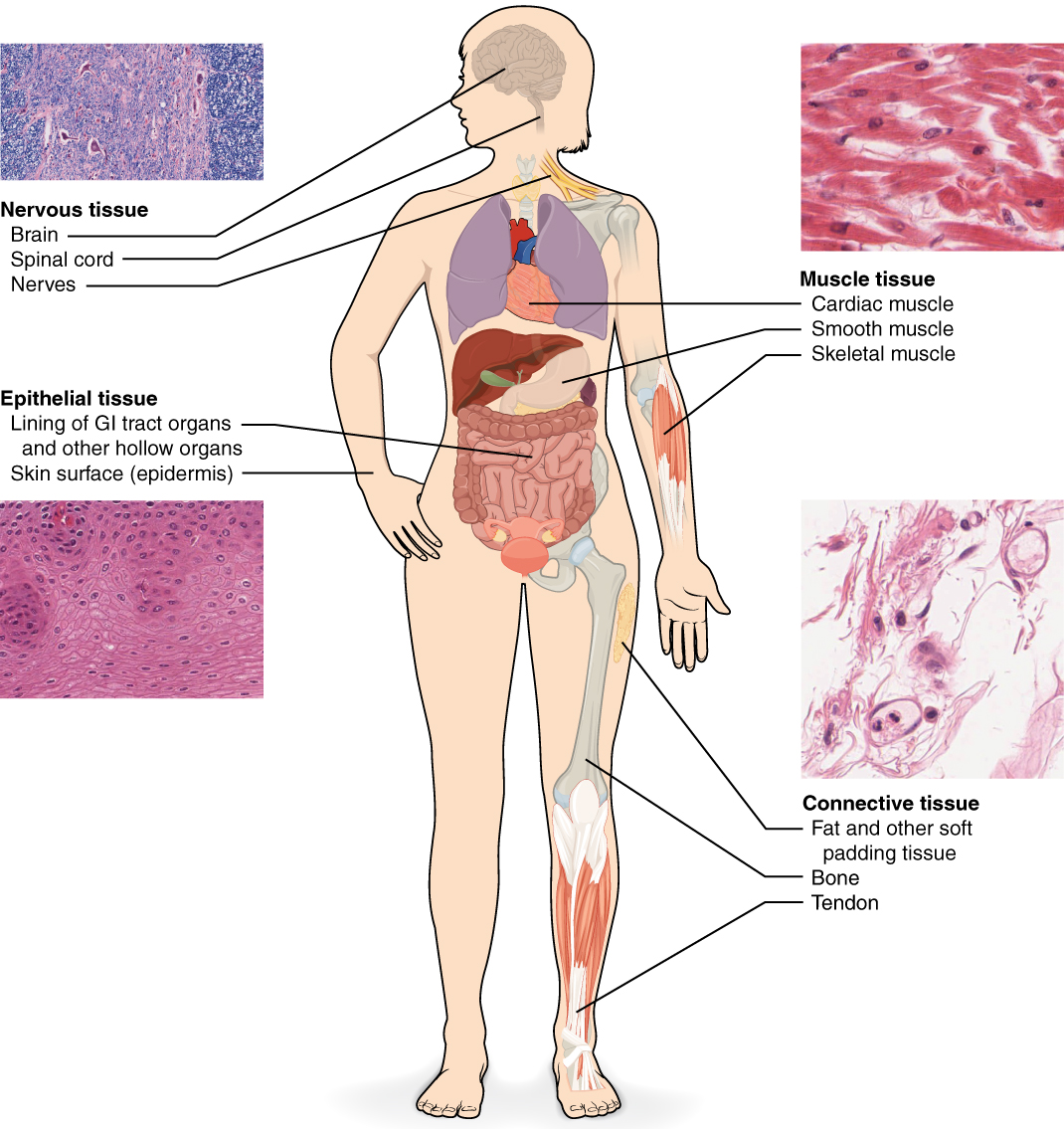
El tejido epitelial, también conocido como epitelio, se refiere a las láminas de células que cubren las superficies exteriores del cuerpo, recubren las cavidades internas y los pasillos, y forman ciertas glándulas. El tejido conectivo, como su nombre lo indica, une las células y órganos del cuerpo y funciona en la protección, soporte e integración de todas las partes del cuerpo. El tejido muscular es excitable, responde a la estimulación y la contraccion para proporcionar movimiento, y se presenta como tres tipos principales: músculo esquelético (voluntario), músculo liso y músculo cardíaco en el corazón. El tejido nervioso también es excitable, permitiendo la propagación de señales electroquímicas en forma de impulsos nerviosos que se comunican entre diferentes regiones del cuerpo (Figura\(\PageIndex{1}\)).
El siguiente nivel de organización es el órgano, donde varios tipos de tejidos se unen para formar una unidad de trabajo. Así como conocer la estructura y función de las células te ayuda en tu estudio de los tejidos, el conocimiento de los tejidos te ayudará a entender cómo funcionan los órganos. Los tejidos epiteliales y conectivos se discuten en detalle en este capítulo. Los tejidos musculares y nerviosos serán discutidos sólo brevemente en este capítulo.
Origen Embrionario de los Tejidos
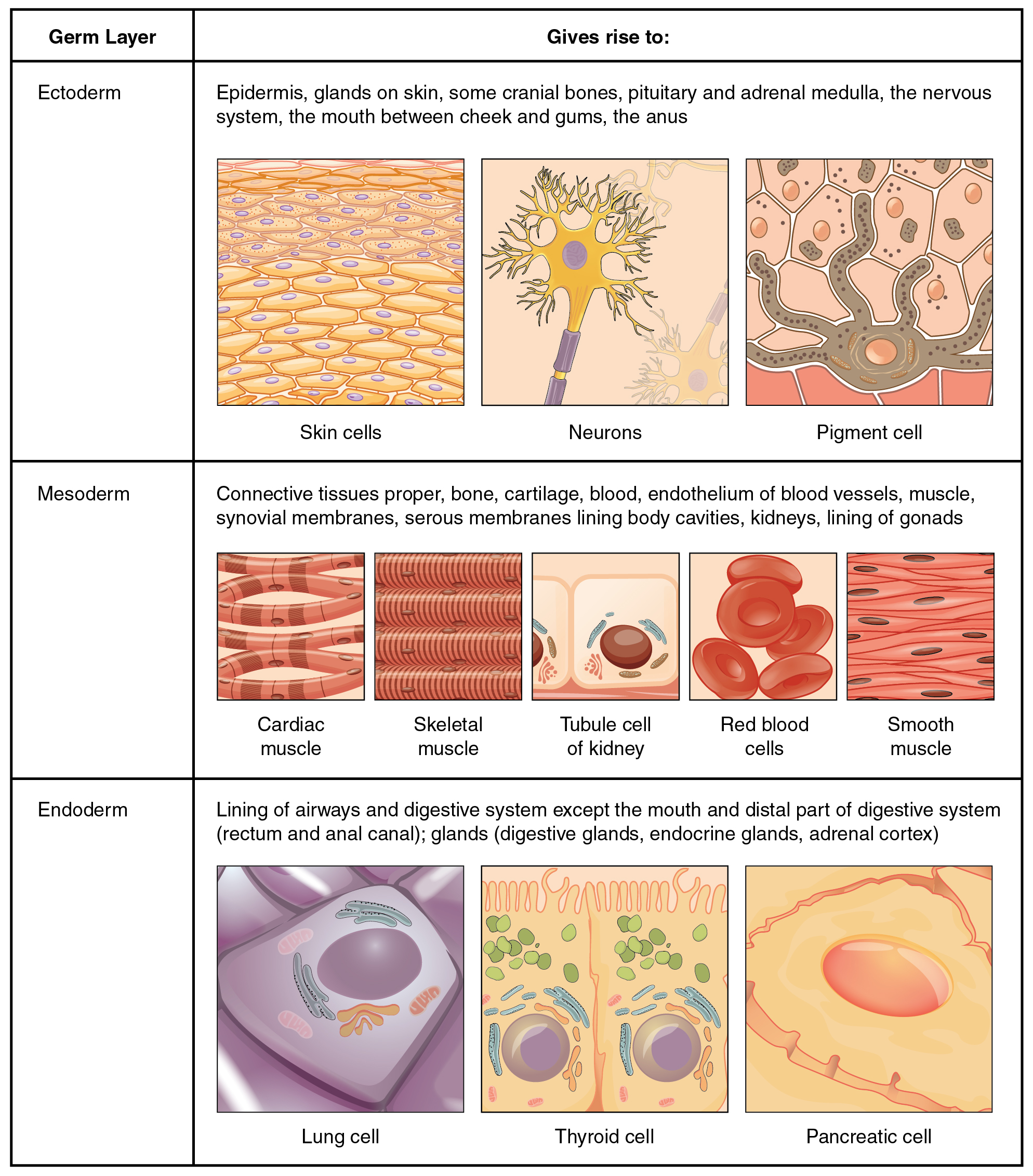
El cigoto, o óvulo fertilizado, es una sola célula formada por la fusión de un óvulo y esperma. Después de la fertilización el cigoto da lugar a rápidos ciclos mitóticos, generando muchas células para formar el embrión. Las primeras células embrionarias generadas tienen la capacidad de diferenciarse en cualquier tipo de célula en el cuerpo y, como tales, se llaman totipotentes, lo que significa que cada una tiene la capacidad de dividirse, diferenciarse y desarrollarse en un nuevo organismo. A medida que avanza la proliferación celular, se establecen tres linajes celulares principales dentro del embrión. Cada uno de estos linajes de células embrionarias forma las distintas capas germinales a partir de las cuales finalmente se forman todos los tejidos y órganos del cuerpo humano. Cada capa germinal se identifica por su posición relativa: ectodermo (ecto- = “exterior”), mesodermo (meso- = “medio”) y endodermo (endo- = “interior”). La figura\(\PageIndex{2}\) muestra los tipos de tejidos y órganos asociados con cada una de las tres capas germinales. Nótese que el tejido epitelial se origina en las tres capas, mientras que el tejido nervioso deriva principalmente del ectodermo y del tejido muscular del mesodermo.

Vea esta presentación de diapositivas para obtener más información sobre las células madre. ¿En qué se diferencian las células madre somáticas de las células madre embrionarias?
Membranas de tejido
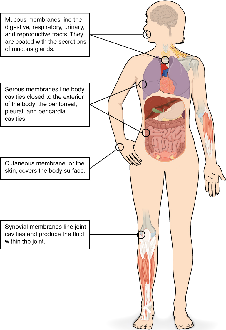
Una membrana tisular es una capa delgada o lámina de células que cubre el exterior del cuerpo (por ejemplo, la piel), los órganos (por ejemplo, el pericardio), los conductos internos que conducen al exterior del cuerpo (por ejemplo, mesenterios abdominales) y el revestimiento de las cavidades articulares móviles. Existen dos tipos básicos de membranas tisulares: tejido conectivo y membranas epiteliales (Figura\(\PageIndex{3}\)).
Connective Tissue Membranes
The connective tissue membrane is formed solely from connective tissue. These membranes encapsulate organs, such as the kidneys, and line our movable joints. A synovial membrane is a type of connective tissue membrane that lines the cavity of a freely movable joint. For example, synovial membranes surround the joints of the shoulder, elbow, and knee. Fibroblasts in the inner layer of the synovial membrane release hyaluronan into the joint cavity. The hyaluronan effectively traps available water to form the synovial fluid, a natural lubricant that enables the bones of a joint to move freely against one another without much friction. This synovial fluid readily exchanges water and nutrients with blood, as do all body fluids.
Epithelial Membranes
The epithelial membrane is composed of epithelium attached to a layer of connective tissue, for example, your skin. The mucous membrane is also a composite of connective and epithelial tissues. Sometimes called mucosae, these epithelial membranes line the body cavities and hollow passageways that open to the external environment, and include the digestive, respiratory, excretory, and reproductive tracts. Mucous, produced by the epithelial exocrine glands, covers the epithelial layer. The underlying connective tissue, called the lamina propria (literally “own layer”), help support the fragile epithelial layer.
A serous membrane is an epithelial membrane composed of mesodermally derived epithelium called the mesothelium that is supported by connective tissue. These membranes line the coelomic cavities of the body, that is, those cavities that do not open to the outside, and they cover the organs located within those cavities. They are essentially membranous bags, with mesothelium lining the inside and connective tissue on the outside. Serous fluid secreted by the cells of the thin squamous mesothelium lubricates the membrane and reduces abrasion and friction between organs. Serous membranes are identified according locations. Three serous membranes line the thoracic cavity; the two pleura that cover the lungs and the pericardium that covers the heart. A fourth, the peritoneum, is the serous membrane in the abdominal cavity that covers abdominal organs and forms double sheets of mesenteries that suspend many of the digestive organs.
The skin is an epithelial membrane also called the cutaneous membrane. It is a stratified squamous epithelial membrane resting on top of connective tissue. The apical surface of this membrane is exposed to the external environment and is covered with dead, keratinized cells that help protect the body from desiccation and pathogens.
Chapter Review
The human body contains more than 200 types of cells that can all be classified into four types of tissues: epithelial, connective, muscle, and nervous. Epithelial tissues act as coverings controlling the movement of materials across the surface. Connective tissue integrates the various parts of the body and provides support and protection to organs. Muscle tissue allows the body to move. Nervous tissues propagate information.
The study of the shape and arrangement of cells in tissue is called histology. All cells and tissues in the body derive from three germ layers in the embryo: the ectoderm, mesoderm, and endoderm.
Different types of tissues form membranes that enclose organs, provide a friction-free interaction between organs, and keep organs together. Synovial membranes are connective tissue membranes that protect and line the joints. Epithelial membranes are formed from epithelial tissue attached to a layer of connective tissue. There are three types of epithelial membranes: mucous, which contain glands; serous, which secrete fluid; and cutaneous which makes up the skin.
Interactive Link Questions
View this slideshow to learn more about stem cells. How do somatic stem cells differ from embryonic stem cells?
Answer: Most somatic stem cells give rise to only a few cell types.
Review Questions
Q. Which of the following is not a type of tissue?
A. muscle
B. nervous
C. embryonic
D. epithelial
Answer: C
Q. The process by which a less specialized cell matures into a more specialized cell is called ________.
A. differentiation
B. maturation
C. modification
D. specialization
Answer: A
Q. Differentiated cells in a developing embryo derive from ________.
A. endothelium, mesothelium, and epithelium
B. ectoderm, mesoderm, and endoderm
C. connective tissue, epithelial tissue, and muscle tissue
D. epidermis, mesoderm, and endothelium
Answer: B
Q. Which of the following lines the body cavities exposed to the external environment?
A. mesothelium
B. lamina propria
C. mesenteries
D. mucosa
Answer: D
Critical Thinking Questions
Q. Identify the four types of tissue in the body, and describe the major functions of each tissue.
A. The four types of tissue in the body are epithelial, connective, muscle, and nervous. Epithelial tissue is made of layers of cells that cover the surfaces of the body that come into contact with the exterior world, line internal cavities, and form glands. Connective tissue binds the cells and organs of the body together and performs many functions, especially in the protection, support, and integration of the body. Muscle tissue, which responds to stimulation and contracts to provide movement, is divided into three major types: skeletal (voluntary) muscles, smooth muscles, and the cardiac muscle in the heart. Nervous tissue allows the body to receive signals and transmit information as electric impulses from one region of the body to another.
Q. The zygote is described as totipotent because it ultimately gives rise to all the cells in your body including the highly specialized cells of your nervous system. Describe this transition, discussing the steps and processes that lead to these specialized cells.
A. The zygote divides into many cells. As these cells become specialized, they lose their ability to differentiate into all tissues. At first they form the three primary germ layers. Following the cells of the ectodermal germ layer, they too become more restricted in what they can form. Ultimately, some of these ectodermal cells become further restricted and differentiate in to nerve cells.
Q. What is the function of synovial membranes?
A. Synovial membranes are a type of connective tissue membrane that supports mobility in joints. The membrane lines the joint cavity and contains fibroblasts that produce hyaluronan, which leads to the production of synovial fluid, a natural lubricant that enables the bones of a joint to move freely against one another.
Glossary
- connective tissue
- type of tissue that serves to hold in place, connect, and integrate the body’s organs and systems
- connective tissue membrane
- connective tissue that encapsulates organs and lines movable joints
- cutaneous membrane
- skin; epithelial tissue made up of a stratified squamous epithelial cells that cover the outside of the body
- ectoderm
- outermost embryonic germ layer from which the epidermis and the nervous tissue derive
- endoderm
- innermost embryonic germ layer from which most of the digestive system and lower respiratory system derive
- epithelial membrane
- epithelium attached to a layer of connective tissue
- epithelial tissue
- type of tissue that serves primarily as a covering or lining of body parts, protecting the body; it also functions in absorption, transport, and secretion
- histology
- microscopic study of tissue architecture, organization, and function
- lamina propria
- areolar connective tissue underlying a mucous membrane
- mesoderm
- middle embryonic germ layer from which connective tissue, muscle tissue, and some epithelial tissue derive
- mucous membrane
- tissue membrane that is covered by protective mucous and lines tissue exposed to the outside environment
- muscle tissue
- type of tissue that is capable of contracting and generating tension in response to stimulation; produces movement.
- nervous tissue
- type of tissue that is capable of sending and receiving impulses through electrochemical signals.
- serous membrane
- type of tissue membrane that lines body cavities and lubricates them with serous fluid
- synovial membrane
- connective tissue membrane that lines the cavities of freely movable joints, producing synovial fluid for lubrication
- tissue
- group of cells that are similar in form and perform related functions
- tissue membrane
- thin layer or sheet of cells that covers the outside of the body, organs, and internal cavities
- totipotent
- embryonic cells that have the ability to differentiate into any type of cell and organ in the body