
25.2: Anatomía macroscópica del transporte de orina
- Page ID
- 123021

\( \newcommand{\vecs}[1]{\overset { \scriptstyle \rightharpoonup} {\mathbf{#1}} } \)
\( \newcommand{\vecd}[1]{\overset{-\!-\!\rightharpoonup}{\vphantom{a}\smash {#1}}} \)
\( \newcommand{\id}{\mathrm{id}}\) \( \newcommand{\Span}{\mathrm{span}}\)
( \newcommand{\kernel}{\mathrm{null}\,}\) \( \newcommand{\range}{\mathrm{range}\,}\)
\( \newcommand{\RealPart}{\mathrm{Re}}\) \( \newcommand{\ImaginaryPart}{\mathrm{Im}}\)
\( \newcommand{\Argument}{\mathrm{Arg}}\) \( \newcommand{\norm}[1]{\| #1 \|}\)
\( \newcommand{\inner}[2]{\langle #1, #2 \rangle}\)
\( \newcommand{\Span}{\mathrm{span}}\)
\( \newcommand{\id}{\mathrm{id}}\)
\( \newcommand{\Span}{\mathrm{span}}\)
\( \newcommand{\kernel}{\mathrm{null}\,}\)
\( \newcommand{\range}{\mathrm{range}\,}\)
\( \newcommand{\RealPart}{\mathrm{Re}}\)
\( \newcommand{\ImaginaryPart}{\mathrm{Im}}\)
\( \newcommand{\Argument}{\mathrm{Arg}}\)
\( \newcommand{\norm}[1]{\| #1 \|}\)
\( \newcommand{\inner}[2]{\langle #1, #2 \rangle}\)
\( \newcommand{\Span}{\mathrm{span}}\) \( \newcommand{\AA}{\unicode[.8,0]{x212B}}\)
\( \newcommand{\vectorA}[1]{\vec{#1}} % arrow\)
\( \newcommand{\vectorAt}[1]{\vec{\text{#1}}} % arrow\)
\( \newcommand{\vectorB}[1]{\overset { \scriptstyle \rightharpoonup} {\mathbf{#1}} } \)
\( \newcommand{\vectorC}[1]{\textbf{#1}} \)
\( \newcommand{\vectorD}[1]{\overrightarrow{#1}} \)
\( \newcommand{\vectorDt}[1]{\overrightarrow{\text{#1}}} \)
\( \newcommand{\vectE}[1]{\overset{-\!-\!\rightharpoonup}{\vphantom{a}\smash{\mathbf {#1}}}} \)
\( \newcommand{\vecs}[1]{\overset { \scriptstyle \rightharpoonup} {\mathbf{#1}} } \)
\( \newcommand{\vecd}[1]{\overset{-\!-\!\rightharpoonup}{\vphantom{a}\smash {#1}}} \)
Objetivos de aprendizaje
- Identificar los uréteres, la vejiga urinaria y la uretra, así como su ubicación, estructura, histología y función
- Comparar y contrastar uretra masculina y femenina
- Describir el reflejo de micción
- Describir el control neuronal voluntario e involuntario de la micción
En lugar de comenzar con la formación de orina, esta sección comenzará con la excreción de orina. La orina es un fluido de composición variable que requiere estructuras especializadas para extraerla del cuerpo de manera segura y eficiente. La sangre se filtra y el filtrado se transforma en orina a una velocidad relativamente constante a lo largo del día. Este líquido procesado se almacena hasta un momento conveniente para su excreción. Todas las estructuras involucradas en el transporte y almacenamiento de la orina son lo suficientemente grandes como para ser visibles a simple vista. Este sistema de transporte y almacenamiento no sólo almacena los desechos, sino que protege los tejidos de daños debido al amplio rango de pH y osmolaridad de la orina, previene la infección por organismos extraños, y para el macho, proporciona funciones reproductivas.
Uretra
La uretra transporta la orina desde la vejiga hacia el exterior del cuerpo para su eliminación. La uretra es el único órgano urológico que muestra alguna diferencia anatómica significativa entre hombres y mujeres; todas las demás estructuras de transporte de orina son idénticas (Figura\(\PageIndex{1}\)).
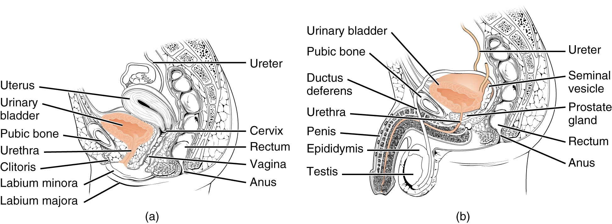
La uretra tanto en machos como en hembras comienza inferior y central a las dos aberturas ureterales formando los tres puntos de una zona triangular en la base de la vejiga llamada trígono (griego tri- = “triángulo” y raíz de la palabra “trigonometría”). La uretra rastrea posterior e inferior a la sínfisis púbica (ver Figura\(\PageIndex{1}\) .a). Tanto en machos como en hembras, la uretra proximal está revestida por epitelio transicional, mientras que la porción terminal es un epitelio escamoso estratificado no queratinizado. En el macho, el epitelio columnar pseudoestratificado recubre la uretra entre estos dos tipos celulares. La micción está regulada por un esfínter urinario interno involuntario controlado por el sistema nervioso autónomo, que consiste en músculo liso y músculo esquelético voluntario que forma el esfínter urinario externo debajo de él.
Uretra Femenina
El orificio uretral externo está incrustado en la pared vaginal anterior inferior al clítoris, superior a la abertura vaginal (introito) y medial a los labios menores. Su corta longitud, cerca de 4 cm, es menos barrera a las bacterias fecales que la uretra masculina más larga y la mejor explicación para la mayor incidencia de ITU en mujeres. El control voluntario del esfínter uretral externo es una función del nervio pudendo. Surge en la región sacra de la médula espinal, viajando a través de los nervios S2-S4 del plexo sacro.
Uretra Masculina
La uretra masculina pasa por la glándula prostática inmediatamente inferior a la vejiga antes de pasar por debajo de la sínfisis púbica (ver Figura\(\PageIndex{1}\) .b). La longitud de la uretra masculina varía entre los hombres pero tiene un promedio de 20 cm de longitud. Se divide en cuatro regiones: la uretra preprostática, la uretra prostática, la uretra membranosa y la uretra esponjosa o peneana. La uretra preprostática es muy corta e incorporada a la pared vesical. La uretra prostática pasa a través de la glándula prostática. Durante las relaciones sexuales, recibe esperma a través de los conductos eyaculatorios y secreciones de las vesículas seminales. Las glándulas de Cowper pareadas (glándulas bulbouretrales) producen y secretan moco en la uretra para amortiguar el pH uretral durante la estimulación sexual. El moco neutraliza el ambiente generalmente ácido y lubrica la uretra, disminuyendo la resistencia a la eyaculación. La uretra membranosa pasa a través de los músculos profundos del perineo, donde es invertida por los esfínteres uretrales suprayacentes. La uretra esponjosa sale por la punta (orificio uretral externo) del pene después de pasar por el cuerpo espongioso. Las glándulas mucosas se encuentran a lo largo de gran parte de la longitud de la uretra y protegen la uretra de los extremos del pH de la orina. La inervación es la misma tanto en machos como en hembras.
Vejiga
La vejiga urinaria recoge orina de ambos uréteres (Figura\(\PageIndex{2}\)). La vejiga se encuentra anterior al útero en las hembras, posterior al hueso púbico y anterior al recto. Durante el embarazo tardío, su capacidad se reduce debido a la compresión por el útero agrandado, lo que resulta en una mayor frecuencia de micción. En los machos, la anatomía es similar, menos el útero, y con la adición de la próstata inferior a la vejiga. La vejiga es parcialmente retroperitoneal (fuera de la cavidad peritoneal) con su “cúpula” cubierta peritoneal que se proyecta hacia el abdomen cuando la vejiga está distendida con orina.
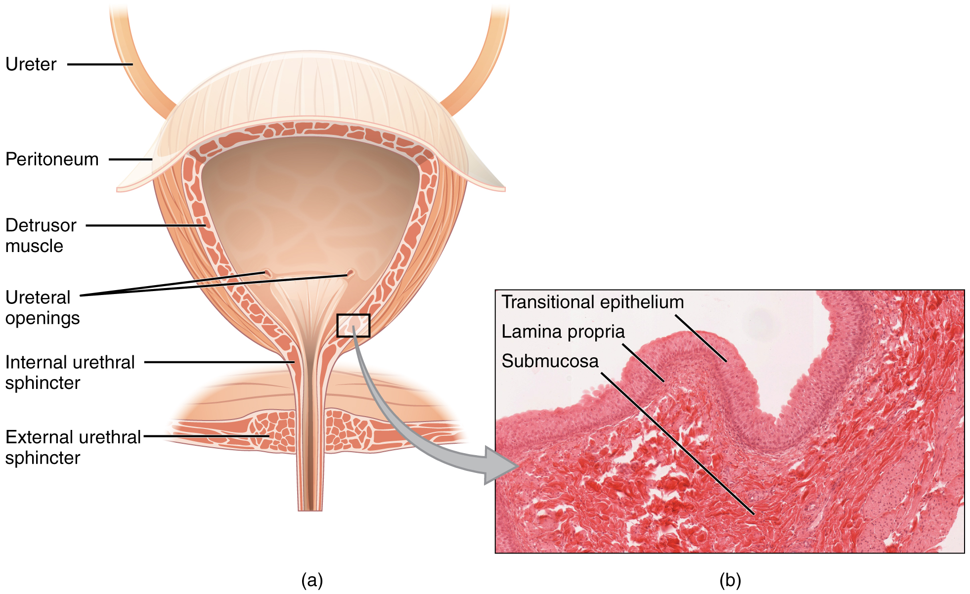

La vejiga es un órgano altamente distensible compuesto por bandas entrecruzadas irregulares de músculo liso llamadas colectivamente músculo detrusor. La superficie interior está hecha de epitelio celular transicional que es estructuralmente adecuado para las grandes fluctuaciones de volumen de la vejiga. Cuando está vacío, se asemeja a epitelios columnares, pero cuando se estira, “pasa” (de ahí el nombre) a una apariencia escamosa (ver Figura\(\PageIndex{2}\)). Volumes in adults can range from nearly zero to 500–600 mL.
The detrusor muscle contracts with significant force in the young. The bladder’s strength diminishes with age, but voluntary contractions of abdominal skeletal muscles can increase intra-abdominal pressure to promote more forceful bladder emptying. Such voluntary contraction is also used in forceful defecation and childbirth.
Micturition Reflex
Micturition is a less-often used, but proper term for urination or voiding. It results from an interplay of involuntary and voluntary actions by the internal and external urethral sphincters. When bladder volume reaches about 150 mL, an urge to void is sensed but is easily overridden. Voluntary control of urination relies on consciously preventing relaxation of the external urethral sphincter to maintain urinary continence. As the bladder fills, subsequent urges become harder to ignore. Ultimately, voluntary constraint fails with resulting incontinence, which will occur as bladder volume approaches 300 to 400 mL.
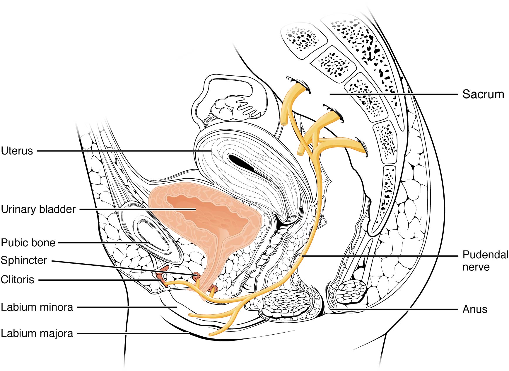
Normal micturition is a result of stretch receptors in the bladder wall that transmit nerve impulses to the sacral region of the spinal cord to generate a spinal reflex. The resulting parasympathetic neural outflow causes contraction of the detrusor muscle and relaxation of the involuntary internal urethral sphincter. At the same time, the spinal cord inhibits somatic motor neurons, resulting in the relaxation of the skeletal muscle of the external urethral sphincter. The micturition reflex is active in infants but with maturity, children learn to override the reflex by asserting external sphincter control, thereby delaying voiding (potty training). This reflex may be preserved even in the face of spinal cord injury that results in paraplegia or quadriplegia. However, relaxation of the external sphincter may not be possible in all cases, and therefore, periodic catheterization may be necessary for bladder emptying.
Nerves involved in the control of urination include the hypogastric, pelvic, and pudendal (Figure \(\PageIndex{3}\)). Voluntary micturition requires an intact spinal cord and functional pudendal nerve arising from the sacral micturition center. Since the external urinary sphincter is voluntary skeletal muscle, actions by cholinergic neurons maintain contraction (and thereby continence) during filling of the bladder. At the same time, sympathetic nervous activity via the hypogastric nerves suppresses contraction of the detrusor muscle. With further bladder stretch, afferent signals traveling over sacral pelvic nerves activate parasympathetic neurons. This activates efferent neurons to release acetylcholine at the neuromuscular junctions, producing detrusor contraction and bladder emptying.
Ureters
The kidneys and ureters are completely retroperitoneal, and the bladder has a peritoneal covering only over the dome. As urine is formed, it drains into the calyces of the kidney, which merge to form the funnel-shaped renal pelvis in the hilum of each kidney. The hilum narrows to become the ureter of each kidney. As urine passes through the ureter, it does not passively drain into the bladder but rather is propelled by waves of peristalsis. As the ureters enter the pelvis, they sweep laterally, hugging the pelvic walls. As they approach the bladder, they turn medially and pierce the bladder wall obliquely. This is important because it creates an one-way valve (a physiological sphincter rather than an anatomical sphincter) that allows urine into the bladder but prevents reflux of urine from the bladder back into the ureter. Children born lacking this oblique course of the ureter through the bladder wall are susceptible to “vesicoureteral reflux,” which dramatically increases their risk of serious UTI. Pregnancy also increases the likelihood of reflux and UTI.
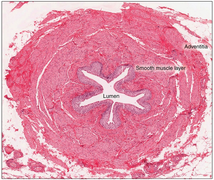
The ureters are approximately 30 cm long. The inner mucosa is lined with transitional epithelium (Figure \(\PageIndex{4}\)) and scattered goblet cells that secrete protective mucus. The muscular layer of the ureter consists of longitudinal and circular smooth muscles that create the peristaltic contractions to move the urine into the bladder without the aid of gravity. Finally, a loose adventitial layer composed of collagen and fat anchors the ureters between the parietal peritoneum and the posterior abdominal wall.
Chapter Review
The urethra is the only urinary structure that differs significantly between males and females. This is due to the dual role of the male urethra in transporting both urine and semen. The urethra arises from the trigone area at the base of the bladder. Urination is controlled by an involuntary internal sphincter of smooth muscle and a voluntary external sphincter of skeletal muscle. The shorter female urethra contributes to the higher incidence of bladder infections in females. The male urethra receives secretions from the prostate gland, Cowper’s gland, and seminal vesicles as well as sperm. The bladder is largely retroperitoneal and can hold up to 500–600 mL urine. Micturition is the process of voiding the urine and involves both involuntary and voluntary actions. Voluntary control of micturition requires a mature and intact sacral micturition center. It also requires an intact spinal cord. Loss of control of micturition is called incontinence and results in voiding when the bladder contains about 250 mL urine. The ureters are retroperitoneal and lead from the renal pelvis of the kidney to the trigone area at the base of the bladder. A thick muscular wall consisting of longitudinal and circular smooth muscle helps move urine toward the bladder by way of peristaltic contractions.
Review Questions
Q. Peristaltic contractions occur in the ________.
A. urethra
B. bladder
C. ureters
D. urethra, bladder, and ureters
Answer: C
Q. Somatic motor neurons must be ________ to relax the external urethral sphincter to allow urination.
A. stimulated
B. inhibited
Answer: B
Q. Which part of the urinary system is not completely retroperitoneal?
A. kidneys
B. ureters
C. bladder
D. nephrons
Answer: C
Critical Thinking Questions
Q. Why are females more likely to contract bladder infections than males?
A. The longer urethra of males means bacteria must travel farther to the bladder to cause an infection.
Q. Describe how forceful urination is accomplished.
A. Forceful urination is accomplished by contraction of abdominal muscles.
Glossary
- anatomical sphincter
- smooth or skeletal muscle surrounding the lumen of a vessel or hollow organ that can restrict flow when contracted
- detrusor muscle
- smooth muscle in the bladder wall; fibers run in all directions to reduce the size of the organ when emptying it of urine
- external urinary sphincter
- skeletal muscle; must be relaxed consciously to void urine
- internal urinary sphincter
- smooth muscle at the juncture of the bladder and urethra; relaxes as the bladder fills to allow urine into the urethra
- incontinence
- loss of ability to control micturition
- micturition
- also called urination or voiding
- physiological sphincter
- sphincter consisting of circular smooth muscle indistinguishable from adjacent muscle but possessing differential innervations, permitting its function as a sphincter; structurally weak
- retroperitoneal
- outside the peritoneal cavity; in the case of the kidney and ureters, between the parietal peritoneum and the abdominal wall
- sacral micturition center
- group of neurons in the sacral region of the spinal cord that controls urination; acts reflexively unless its action is modified by higher brain centers to allow voluntary urination
- trigone
- area at the base of the bladder marked by the two ureters in the posterior–lateral aspect and the urethral orifice in the anterior aspect oriented like points on a triangle
- urethra
- transports urine from the bladder to the outside environment